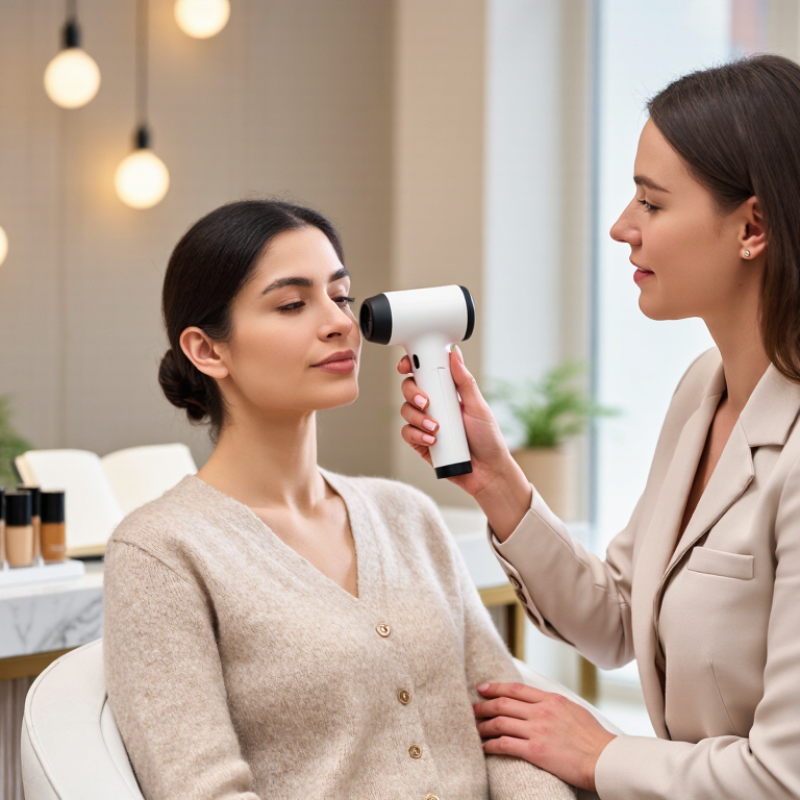
| 1. Reflectance spectrophotometer | Spectral reflectance; may report CIELAB coordinates (L*, a*, b*) and derived color differences. Some systems estimate melanin- or erythema-related indices. | Measures reflected light across multiple wavelengths at a contact or defined measurement area. | Objective color assessment, foundation shade matching, and monitoring color changes under a consistent protocol. | More repeatable than unaided visual judgment when calibrated and used consistently. | Results depend on device geometry, calibration, site selection, and skin preparation; derived indices are not interchangeable across devices. |
| 2. Tristimulus colorimeter | Color values approximating human visual response, commonly expressed as L*, a*, and b* in compatible systems. | Uses filtered sensors to capture a small set of color-response signals rather than a full reflectance spectrum. | Quick, localized complexion measurements and before-and-after comparisons. | Typically compact and straightforward for repeated measurements at the same site. | Provides less spectral detail than a spectrophotometer; readings can be affected by pressure, placement, and ambient conditions. |
| 3. Cross-polarized imaging system | Standardized facial images that can help visualize surface redness, pigmentation contrast, and visible texture patterns. | Uses polarized illumination and imaging to reduce surface glare and capture more consistent-looking photographs. | Consultation records, visual treatment tracking, and identifying areas for closer assessment. | Captures a broad facial area and supports side-by-side image review. | Images are not direct measurements of pigment concentration; lighting, pose, exposure, and software processing affect comparisons. |
| 4. Multispectral imaging system | Images or reflectance information captured in several wavelength bands; software may estimate visible pigment and redness patterns. | Records the skin under multiple selected wavelengths, sometimes including bands beyond visible light. | Detailed complexion consultations and standardized tracking of visible skin features. | Can reveal image patterns that are difficult to distinguish in ordinary photographs. | Software outputs are estimates, not diagnoses; interpretation requires consistent capture conditions and clear disclosure of what the device can validate. |
| 5. Standardized RGB imaging tool | Digital color values and image-based estimates of complexion tone or visible color variation. | Uses a camera with controlled lighting, fixed distance, and, ideally, a color reference for image consistency. | Accessible shade consultation, digital records, and basic visual comparison. | Convenient and easy to incorporate into a photo-based consultation workflow. | Camera sensors, automatic exposure, white balance, and lighting can shift apparent color; uncalibrated RGB values should not be treated as precise skin measurements. |
| 6. Dermatoscopic imaging device | Magnified, illuminated views of localized skin features, including surface patterns and color distribution. | Uses magnification and controlled illumination; some devices use polarization to reduce surface reflection. | Close-up documentation of a specific area when appropriate to the service and operator training. | Provides more localized detail than a standard facial photograph. | Does not measure overall skin tone or establish a diagnosis; suspicious or changing lesions should be referred to a qualified medical professional. |
| 7. Standardized visual assessment | A trained observer's categorical assessment of visible tone or color response using a defined chart or scale. | Compares skin under consistent neutral lighting with a documented scale and assessment site. | Initial consultations, communication about shade ranges, and settings without instrumental equipment. | Low-cost, accessible, and useful when paired with clear terminology and a consistent procedure. | Subject to observer variation, lighting, and surrounding colors; categories are not equivalent to an instrumental measurement. |