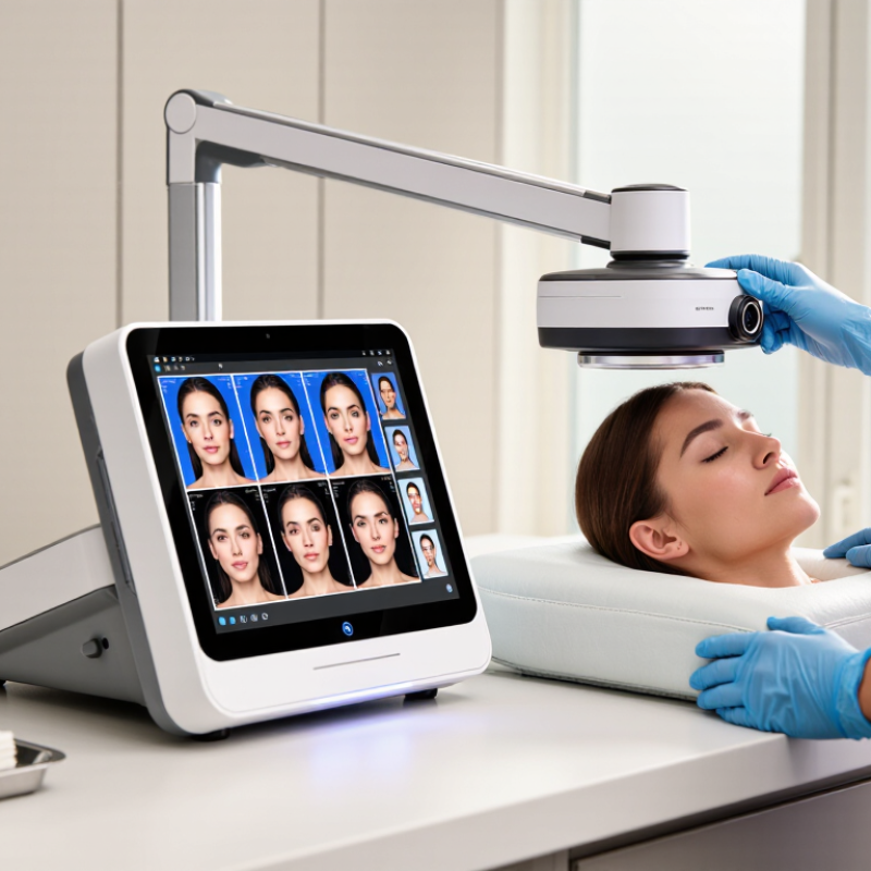
| Standardized visible-light imaging | Color photographs of the skin's visible appearance under controlled illumination. | Baseline documentation; visible redness, spots, uneven tone, and general appearance. | Intuitive images that are easy to review and compare over time. | Results can vary with lighting, camera settings, pose, distance, and white balance. A photograph alone does not establish a diagnosis. | Consistent lighting, fixed positioning, repeatable camera settings, and clear image-storage and comparison tools. |
| Cross-polarized imaging | Images captured with crossed polarizing filters that reduce surface glare and can make some color variations beneath the skin surface easier to see. | Documenting visible pigmentation and redness patterns with less reflection from the skin surface. | Reduces specular shine, which can otherwise obscure details in photographs. | Image appearance depends on the light source, filters, and processing; it does not reveal skin structures with diagnostic certainty. | Whether images are repeatable across sessions and whether the system explains how images are processed. |
| Parallel-polarized imaging | Polarized light and camera filters aligned in the same direction, retaining more surface reflection than cross-polarized imaging. | Visualizing surface texture, fine lines, and some skin-surface irregularities. | Can emphasize surface detail and complement other image modes. | Reflections can vary with skin oil, moisture, product residue, and positioning; findings are image-based estimates. | Image consistency, preparation instructions, and whether the feature adds useful information beyond standard photography. |
| UV-induced fluorescence imaging | Fluorescence emitted by certain skin-surface substances when illuminated with ultraviolet or near-ultraviolet light. | Showing some surface features, such as fluorescence associated with porphyrins, and assisting visual skin assessments. | Provides an image mode not visible in ordinary white-light photographs. | Fluorescence can be affected by cosmetics, cleansers, and other surface substances. It is not a standalone test for acne, infection, or other conditions. | Light wavelength, eye-safety guidance, cleaning and preparation protocol, and the intended interpretation of the images. |
| Multispectral imaging | Images acquired at multiple selected wavelength bands, which can provide different information about light absorption and reflection in skin. | Estimating and mapping features associated with pigmentation, redness, or other optical characteristics. | Offers more wavelength-specific information than a single visible-light image. | Outputs depend on wavelength selection, calibration, algorithms, and skin characteristics. A displayed map is an estimate, not a direct measurement of every skin layer. | Available wavelength bands, calibration procedures, validation information, and clear definitions of each reported score or map. |
| 3D imaging or stereophotogrammetry | A three-dimensional surface model reconstructed from multiple images or views. | Assessing facial surface contours, volume changes, and some topographic features over time. | Adds surface-shape information that a flat photograph cannot provide. | Accuracy depends on capture conditions, registration, facial expression, and movement. It does not show internal tissue directly. | Capture time, repeatability, movement tolerance, measurement units, and tools for aligning follow-up scans. |
| Automated image analysis | Software-generated estimates or scores for image features such as visible spots, pores, wrinkles, or redness. | Organizing observations, supporting consultations, and tracking changes in standardized images. | Can make image review more structured and help compare results across visits. | Scores depend on the algorithm, image quality, and population used for validation; different systems may use non-equivalent scales. | Definitions for each score, validation details, export options, and whether results can be reviewed alongside the original images. |
| Calibration and repeatability | Procedures and design features that help keep image capture and measurements consistent between sessions. | Longitudinal tracking and fair comparison of images or scores over time. | Improves the usefulness of follow-up comparisons when capture conditions are controlled. | Consistency does not by itself prove clinical accuracy or make results comparable across different devices. | Routine calibration needs, fixed positioning aids, standardized protocols, and documented repeatability data. |
| Data management and privacy | How images, measurements, and client or patient records are stored, accessed, exported, and protected. | Maintaining records, reviewing progress, and sharing results with authorized users. | Supports organized follow-up and more efficient image comparison. | Cloud storage, access controls, retention, and data-transfer practices differ; privacy obligations depend on location and use. | Consent workflow, encryption, user permissions, backup and deletion policies, export format, and applicable privacy requirements. |